
| 日本放射光学会誌 Journal of JSSRR |
 |
 |
| Vol.30,No.5/Sept. 2017 |
|
|
 |
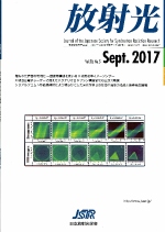
| 【表紙の説明】 X線反射投影による多層膜の可視化。シリコン基板上に金薄膜(厚さ約150Å、三角形パターン)、銅薄膜(厚さ約160Å、均一)、ジルコニウム薄膜(厚さ約240Å、円形パターン)を積層した試料。視射角と面内角の両方を変化させて得たX線反射投影サイノグラム(上図)とその画像再構成演算で求まるX線反射率の試料内分布(下図)。 Visualization of multilayered thin films by X-ray reflection projection.The used sample is Zr(around 240 angstrom, circular pattern)/Cu(around 160 angstrom, uniform)/Au(around 150 angstrom, triangle pattern)/Si(substrate). Top: X-ray reflection projection sinogram obtained by scanning both grazing and in-plane angles. Bottom: X-ray reflectivity distribution in the sample, which is obtained from image reconstruction of the sinogram. |
| ・ |  |
| *放射光の発展と共に歩んだ筋収縮研究の50年と構造生物学 若林克三(p.209)(2ページ、936k) |
|
| ・ |  |
| *埋もれた界面の可視化 -画像再構成を用いるX線反射率イメージング- 桜井健次、蒋 金星、平野馨一(p.211) *Visualization of buried interfaces:X-ray reflectivity imaging using image reconstruction Kenji SAKURAI, Jinxing JIANG and Keiichi HIRANO (7 pages, 2,991k) |
|
| *X線自由電子レーザーで捉えたバクテリオロドプシン構造変化の三次元動画 南後恵理子、久保 稔、岩田 想(p.218) *A three-dimensional movie of structural changes in bacteriorhodopsin captured by X-ray free electron lasers Eriko NANGO, Minoru KUBO and So IWATA (10 pages, 3,083k) |
|
| *シリアルフェムト秒結晶解析により明らかにした光化学系IIの反応中間体の構造と酸素発生機構 菅 倫寛、秋田総理、菅原道泰、久保 稔、岩田 想、沈 建仁(p.228) *Intermediate structure and oxygen-evolving mechanism of photosystem II revealed by serial femtosecond crystallography Michihiro SUGA, Fusamichi AKITA, Michihiro SUGAHARA, Minoru KUBO, So IWATA and Jian-Ren SHEN (7 pages, 2,335k) |
|
| ・ |  |
| *中東放射光施設 SESAME 野村昌治(p.235) (3 pages, 2,641k) |
|
| *The 1st Asia Oceania Forum Synchrotron Radiation Schoolに参加して 高木秀彰、富田翔伍(p.238) (2 pages, 1,224k) |
|
| *日本放射光学会若手部会設立について 和達大樹、片山哲夫、永村直佳、堀川裕加、山崎裕一、山田悠介(p.240) (2 pages, 1,416k) |
|
| ・ |
| Back |